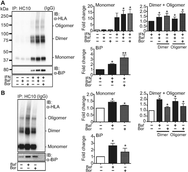
Figure 1.
Accumulation of HLA-B27 with inhibition of autophagy or ERAD. (A) BM-derived macrophages were left untreated or stimulated with IFNγ (IFN) for 21 hours, then treated for 3 hours with bafilomycin (100 nM) (Baf) to block the autophagy pathway, or with bortezomib (10 nM) (Bor) to block ERAD. Untreated cells and cells only treated with bafilomycin or bortezomib were used as controls. Immunoprecipitation (IP) and immunoblotting (IB) with HC10 and 3B10.7 (α-HLA) or α-BiP, were as indicated and described in Materials and Methods. Dimers and oligomers are visualized on non-reduced samples electrophoresed on non-reducing gels, while BiP is analyzed under reducing conditions. For quantification, samples were normalized to untreated or IFNγ-treated controls, as indicated, and expressed as fold change. For all figures, representative immunoblots are shown. Quantitative data represent mean ± SEM of five independent experiments. (*, p < 0.05) (B) HLA-B27 expressing C1R cells were stimulated for 3 hours with bafilomycin (Baf) or bortezomib (Bor), prior to immunoprecipitation and immunoblotting. Quantitative data represent mean ± SEM of six experiments with the same cell line. (*, p < 0.05). IgG lanes show background when IP was performed with non-specific IgG alone.

