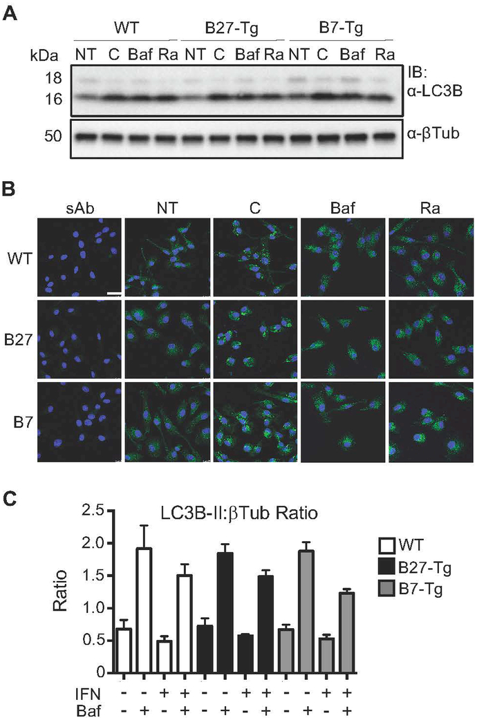
Figure 2.
Autophagy in rat macrophages expressing HLA class I transgenes. (A) BM macrophages from WT, B27-Tg, and B7-Tg rats were left untreated or stimulated with rapamycin (25 μM) (Ra) to induce autophagy, chloroquine (50 μM) (C) or bafilomycin (400 nM) (Baf) to block autophagic flux, and then collected 2 hours later and lysed for immunoblotting using anti-LC3B (α-LC3B), with anti-βTubulin (α-βTub) used as a loading control. Immunoblot shown is representative of three independent experiments. (B) Macrophages treated as described in (A) (NT, untreated; sAb, secondary Ab only) were analyzed by immunofluorescence with anti-LC3B (green), which has a stronger affinity for LC3B-II. Nuclei are visualized by DAPI staining (blue). Immunofluorescence was evaluated using confocal imaging with results shown at 63x magnification. Images are representative of three independent experiments. (C) BM macrophages from WT, B27-Tg, and B7-Tg rats were incubated without or with IFNγ (50 ng/ml) for 24 hours, followed by another 24 hours with bafilomycin (10 nM). LC3B-II expression was determined by immunoblotting (as for experiment shown in A), and is expressed as a ratio of LC3B-II:βTub. Results are representative of three independent experiments.

