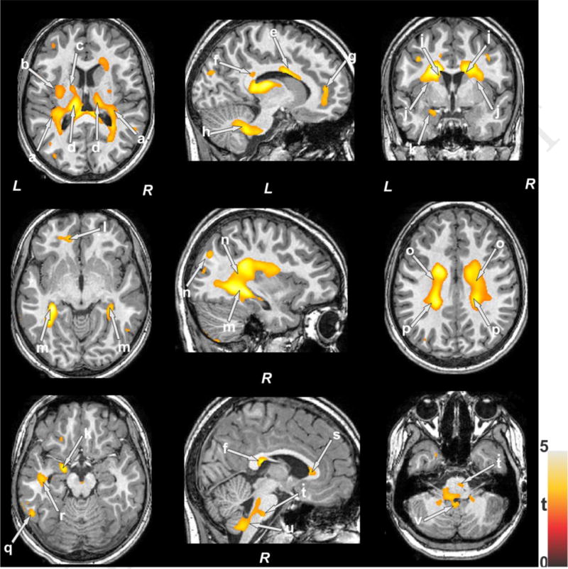
Figure 1.

Brain sites showing lower entropy in OSA compared to control subjects. These sites included the insular cortices (a), external (b) and internal (c) capsules, bilateral anterior, mid, and posterior thalamus (d), anterior (s), mid (e), and posterior (f) corpus callosum, medial prefrontal cortex (g), inferior, middle, and superior cerebellar peduncles (h), bilateral caudate (i), bilateral putamen (j), amygdala (k), prefrontal white matter (k), bilateral hippocampus (m), parietal cortices (n), mid (o) and posterior (p) corona radiate, occipital cortex (q), temporal white matter (r), midline and caudal pons (t), ventral medulla (u), and cerebellar cortices (v). Color bar represents t-statistic values (L = Left; R = Right).
