
Figure 7.
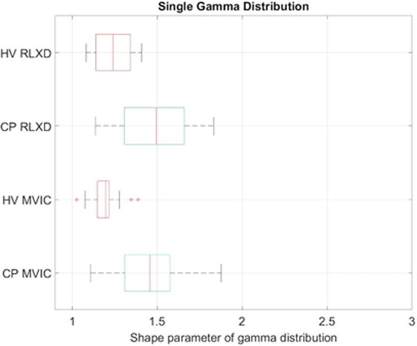
Boxplots of the shape parameter for a single gamma distribution fitted to the speckle envelope in the rectus femoris muscle of healthy volunteers (HV) and subjects with cerebral palsy (CP) when the muscle is relaxed (RLXD) and during maximal voluntary isometric contraction (MVIC). The central line in the boxplot corresponds to the median, the edges correspond to 25th and 75th percentiles, and whiskers correspond to extremal values not considered outliers.