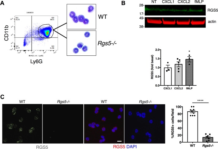
Figure 1.
Immunoreactive RGS5 is detected in mouse neutrophils. A, neutrophils were isolated from bone marrow of naïve mice using Ly6G microbeads. Flow cytometry was used to assess purity based on CD11b expression. Neutrophils were dispersed by cytospin and identified by modified Giemsa staining. The plot is from a single experiment representative of three experiments (purity of 93.2 ± 3%). B, BM-derived neutrophils were left untreated (NT) or stimulated with CXCL1 or CXCL2 (100 ng/ml) or fMLP (1 μm) for 2 h followed by cell lysis and immunoblotting. The bar graph shows the relative RGS5 expression (normalized by β-actin signal; error bars indicate mean ± S.E.) of three to five independent experiments using one to two mice/experiment. *, p = 0.04, one-way ANOVA, Dunnett's post hoc test versus control (unstimulated condition). C, CXCL2-treated neutrophils from WT or Rgs5−/− mice were stained with anti-RGS5 (grayscale in first two images on the left and red in the color images) and counterstained with DAPI to identify nuclei (blue). The bar graph shows the percentage of cells/field containing immunoreactive RGS5 (error bars indicate mean ± S.E. of >3 fields/experiment, each containing >10 cells/field) analyzed in three independent experiments using one mouse of each genotype/experiment. ****, p < 0.0001, unpaired t test. Scale bar, 6 μm.

