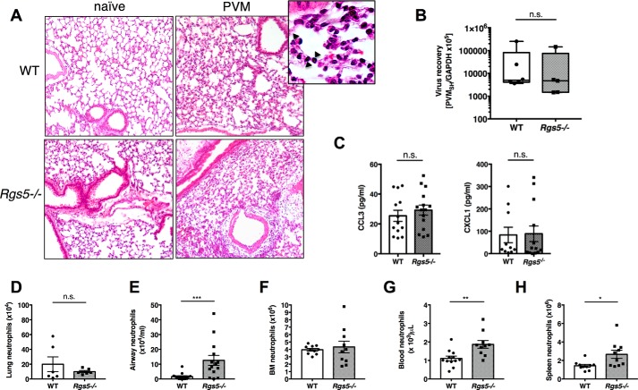
Figure 3.
RGS5 deficiency results increased in airway neutrophils in acute respiratory virus infection. A, hematoxylin and eosin–stained lung sections from WT or Rgs5−/− mice at baseline (naïve) and 5 days after inoculation with PVM. Images are from a single mouse representative of three to four mice/group. Inset, diffuse neutrophilic alveolitis characteristic of acute PVM infection (40× magnification); arrows denote neutrophils. B, virus recovery was assessed by qPCR detection of virus-specific SH gene. C, chemokines CCL3 and CXCL1 in BALF of PVM-infected WT and Rgs5−/− mice. D–G, neutrophils in lung (D), airways (E), bone marrow (F), peripheral blood (G), or spleens (H) at day 5 of PVM infection. *, p = 0.02; **, p = 0.002; ***, p = 0.0007, Mann–Whitney; n.s., not significant. All error bars indicate mean ± S.E.; each symbol represents one mouse.

