Figure 6.
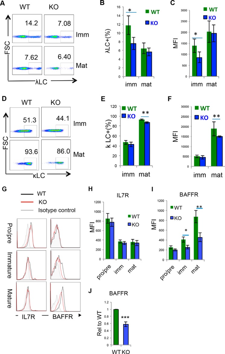
Loss of Hrd1 resulted in the developmental defect of mature B cells. WT and Hrd1 KO (KO) BM cells were isolated and analyzed by flow cytometry. The B220+IgM− pro/pre–B, B220LoIgM+ immature and B220HIIgM+ mature B cells were gated for the analysis. A–F, κLC and λLC expression were analyzed. Representative flow cytometry images for the expression of κLC (A) and λLC (D) in B220LOIgM+ immature B cells and B220HIIgM− mature B cells are shown. Percentages (B) and MFI (C) of κLC expression as analyzed in A. Percentages (E) and MFI (F) of λLC as analyzed in D. G–J, the expression of IL-7R and BAFF receptor (BAFFR) were analyzed. Representative images are shown in G. The MFI of IL-7R (H) and BAFFR (I) from 7 pairs of mice are indicated. The expression levels of BAFFR mRNA in IgM+B220+ cells were determined by real-time RT-PCR (J). Error bars represent S.D. n = 8. *, p < 0.05; **, p < 0.01; ***, p < 0.001.

