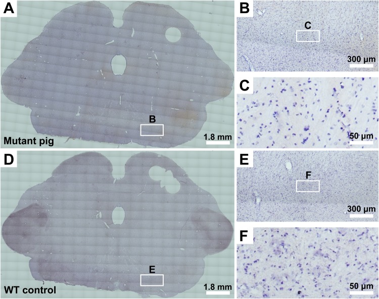
Figure 3.
Detection of Parkinson’s disease (PD)-specific pathological changes in a mutant Guangxi Bama minipig. (A–C) One 3-month-old mutant Guangxi Bama minipig (1–2#) was used to detect PD-specific pathological changes in the substantia nigra by α-synuclein immunohistochemical staining. (D–F) One age-matched wild-type (WT) minipig (1–4#) was used as a negative control. (B,C,E,F) Higher-magnification images show no classical α-synuclein-immunopositive pathology in the substantia nigra of the mutant minipig at 3 months of age (B,C) compared to the age-matched WT minipig (E,F). Note that the immunohistochemistry was performed on the brain tissue from the same two minipigs that were euthanized for mRNA analysis.

