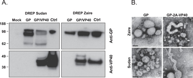
Figure 2.
Analysis of Ebolavirus GP and VP40 expression. (A) BHK-21 cells were transfected with DREP expressing either the GP or GP and VP40 genes from SUDV or EBOV. Cell lysates were collected and run on an SDS-PAGE. Western blotting was performed using anti-GP and anti-VP40 antibodies. EBOV and SUDV VLPs (Ctrl) were used as positive controls. The blots have been cropped and full blots are available in Supplementary Information. (B) Replicon-driven expression of SUDV and EBOV GP and GP-VP40 VLPs was analyzed by electron microscopy (EM) from cell culture supernatants from cells expressing GP and GP-VP40. EM micrographs of VLP preparations reveal pleomorphic VLPs made from GP expressing cells (left panels), and filamentous VLP structures made from GP-VP40 expressing cells (right panels). Black bars indicate 100 nm.

