Figure 1.
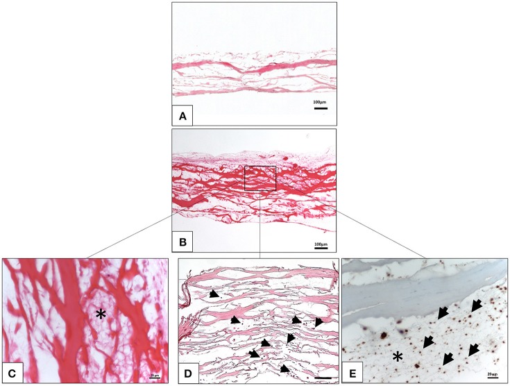
Ex vivo interaction between liquid platelet-rich fibrin and the collagen membrane SB. (A) A control of the SB illustrating the membrane-specific porous structure (H and E staining; x10 magnification; scale bar = 100 μm). (B) Total penetration of leukocytes and platelets from liquid PRF into the SB central region (H and E staining; x100 magnification; scale bar = 100 μm). (C) High magnification micrograph showing the fibrin network (*) within the SB collagen fibers (H and E staining; x400 magnification; scale bar = 20 μm). (D) High magnification micrograph showing the leukocytes (black arrows) within the SB collagen fibers (H and E staining; x200 magnification; scale bar = 100 μm). (E) High magnification micrograph showing the platelets (black arrows) and fibrin network (*) within the SB collagen fibers (anti CD-61 staining; x400 magnification; scale bar = 20 μm).

