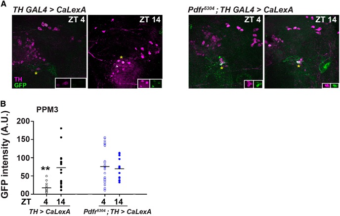
Figure 6.
Intracellular Ca2+ levels in PPM3 neurons lower during day than night but remain similar during day and night in the absence of PDFR. A, TH GAL4 expressing CaLexA in WT and pdfr 5304 backgrounds costained with antibodies against TH to mark dopaminergic neurons and GFP to quantify intracellular Ca2+ levels at two time points: ZT4 and ZT14. CaLexA-driven GFP+ signal was detected at a low level at ZT4 whereas higher intensity at ZT14 (left panels) in WT background. A.U. = arbitrary units. CaLexA-driven GFP+ signal was detected at similar high level at ZT4 and ZT14 (right panels) in pdfr 5304 background. Asterisks indicate PPM3 neurons which are zoomed in inset. B, Quantification of results in A shows significantly lower GFP fluorescence in TH GAL4 > CaLexA flies at ZT4 as compared to GFP fluorescence in TH GAL4 > CaLexA flies at ZT14, pdfr 5304; TH GAL4 > CaLexA flies at ZT4 and ZT14. All other details are as in Figure 1. See also Extended Data Figures 6-1, 6-2.

