Figure 1.
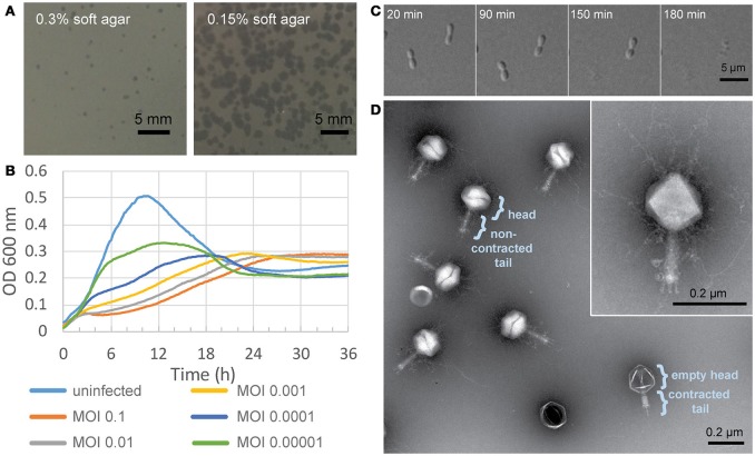
Characterization of Atu_ph07. (A) Atu_ph07 forms small plaques on a lawn of A. tumefaciens C58 on 0.3% soft agar (left) and larger plaques on 0.15% soft agar (right). (B) Growth curve of A. tumefaciens infected with Atu_ph07 at different MOIs. (C) Time-lapse microscopy of A. tumefaciens cells infected with Atu_ph07 at an MOI of 10. (D) TEM image of Atu_ph07 shows the phage is in the family Myoviridae. Inset of a single phage particle at higher magnification is shown to emphasize the presence of tail fibers and whiskers.

