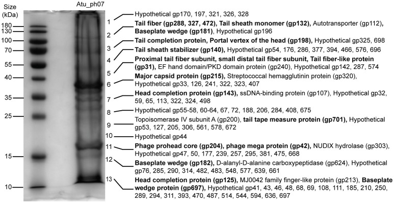
Figure 7.
SDS-PAGE of Atu_ph07 structural proteins as identified by ESI-MS/MS. Phage proteins were separated by size and fragments covering the full lane of the gel were excised for proteomic analysis. Numbers at the right of the gel indicate the position of fragments which were excised from the gel. Proteins identified in each fragment are listed. Bold font indicates validation of annotated structural proteins.

