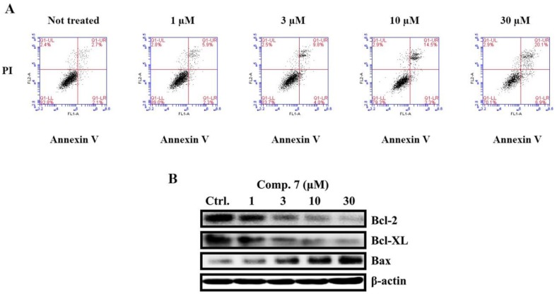
Figure 5.
Apoptosis induced by compound 7 in MCF-7 cells. (A) Apoptosis quantification, using Annexin V/PI double staining assay after treatment with compound 7 for 48 h. MCF-7 cells were harvested and stained with PI and Annexin V-FITC in darkness for 15 min, followed by flow cytometry analysis. Lower right: Annexin V+/PI− (early apoptosis); upper right: Annexin V+/PI+ (late apoptosis and necrosis). (B) The expression of Bcl-2, Bcl-XL and Bax was assessed through western blotting analyses of MCF-7 (treated for 48 h with varying concentrations of the compound), β-actin was used as internal control.

