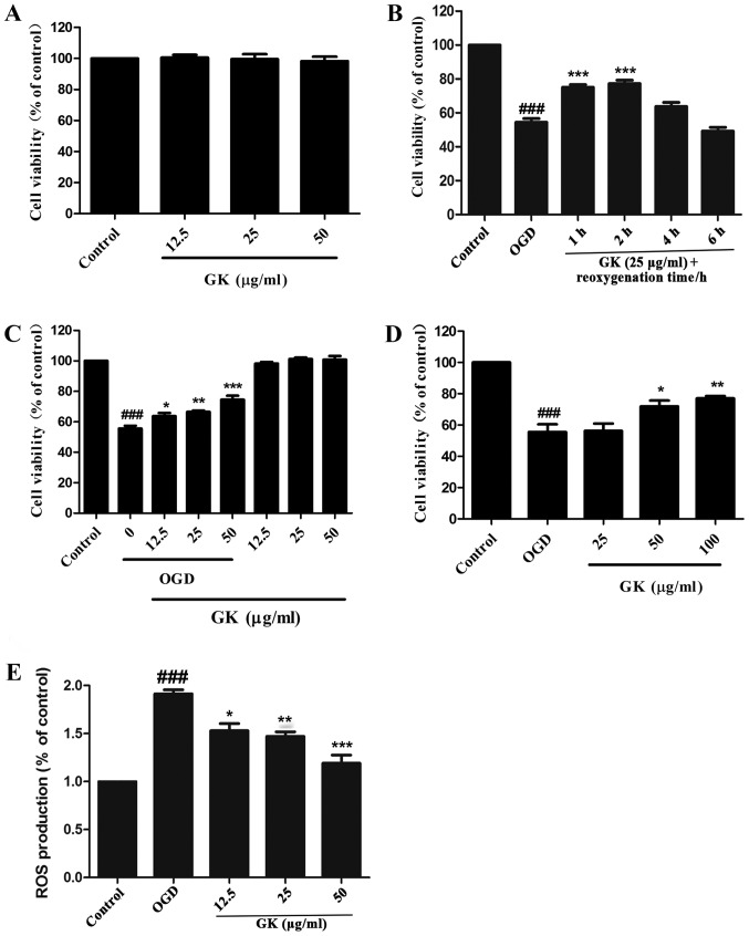
Figure 2.
GK increased cell viability and ROS generation of SH-SY5Y cells damaged by OGD. (A) Cells were treated with 12.5, 25 and 50 µg/ml of GK for 24 h and cell viability was measured with the CCK-8 assay. (B) After 4 h OGD, the SH-SY5Y cells were treated with GK and reoxygenation with various times (1, 2, 4 and 6 h). Cell viability was assessed by CCK-8 assay. (C) After 4 h OGD, the SH-SY5Y cells were treated with GK at different concentrations (12.5, 25 and 50 µg/ml) and reoxygenation for 1 h. Cell viability was assessed by CCK-8 assay. (D) After 4 h OGD, the SH-SY5Y cells were treated with GK at different concentrations (25, 50 and 100 µg/ml) and reoxygenation for 24 h. Cell viability was assessed by CCK-8 assay. (E) ROS generation was quantified by DCFHDA assay at excitation 488 nm and emission 525 nm. Data are represented as mean ± SD from six experiments. ###P<0.001 vs. control group. *P<0.05, **P<0.01 and ***P<0.001 vs. OGD group. GK, ginkgolide K; ROS, reactive oxygen species; CCK-8, Cell Counting Kit-8; OGD, oxygen-glucose deprivation.

