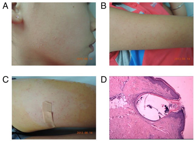
Figure 2.

Clinical and pathological features of keratosis pilaris. (A-C) Clinical images of the proband: Symmetric, rough keratotic follicular papule and mild perifollicular erythema on the cheek, extensor of the upper arms and the anterior thighs. (D) Histopathology of a skin biopsy of the right thigh of the proband revealed that the follicular orifice was distended by a keratin plug; mild infiltration of mononuclear cells in the superficial dermis was observed (hematoxylin and eosin, magnification, ×200).
