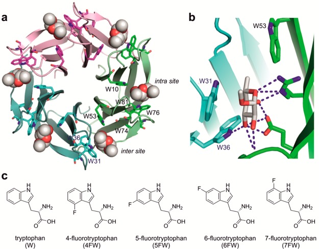
Figure 1.
(a) Structure of RSL (pdb 2BT9). The three monomers are colored in magenta, green, and cyan; the bound αMeFuc is represented as spheres. (b) The intermonomeric binding site with three important Trp residues: W31, W36, and W53 (structurally equivalent to W76, W81, and W10 in the intramonomeric site). (c) Structures of l-tryptophan and the fluorinated l-analogs used in this study.

