Figure 3.

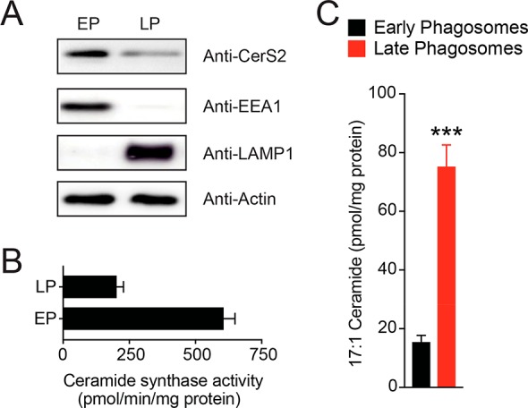
Characterization of CerS2 activity during phagosomal maturation. (A) Western blot analysis confirming the enrichment of CerS2 on EPs. As controls, EEA1 and LAMP1 were used to assess the purity of EP and LP preparations, respectively. In each case, 25 μg of lysate was loaded for all samples in this analysis, and actin was used as a loading control. The Western blot analysis was performed on eight biological replicates with reproducible results. (B) Ceramide synthase in vitro activity assay on EPs and LPs from RAW264.7 mouse macrophages, showing heightened ceramide synthase activity on EPs. Data represents mean ± SEM for three biological replicates per group. (C) Levels of C17:1 containing ceramide on EPs and LPs, following feeding of RAW264.7 mouse macrophages with 1 mM C17:1 FFA (4 h, 37 °C). Data represent mean ± SEM for five biological replicates. ***P < 0.001 for LP group versus EP group by Student’s t test.
