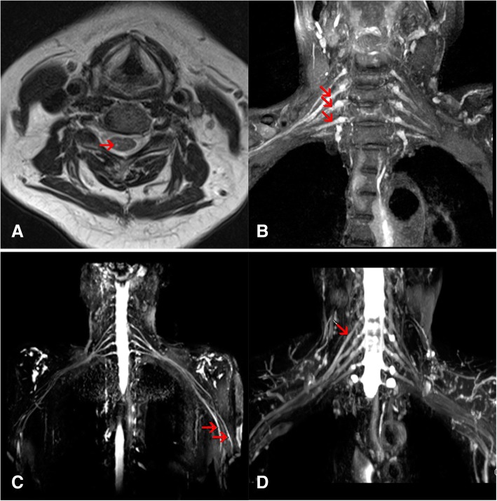
Fig. 4.
Imaging characteristics of patients with SZP. Axial T2-weighted image showed the unilateral hyperintensity in the dorsal horn of C5 spinal cord in patient 2 (a). Brachial plexus magnetic resonance imaging showed hyperintensity of C6–8 nerve roots in patient 5 (b), left median and radial nerves in patient 7 (c) and C5 nerve roots in patient 8 (d)

