. 2017 May 24;8(8):5606–5615. doi: 10.1039/c7sc01837a

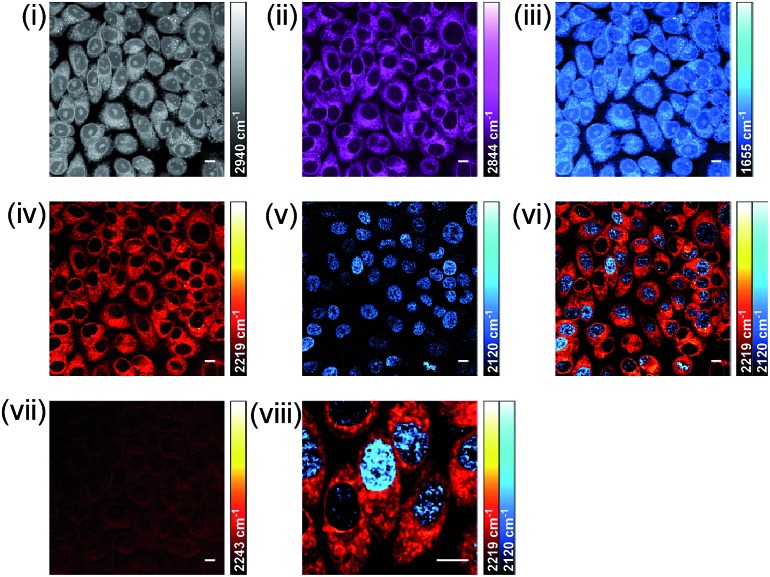
Fig. 8. Dual-colour alkyne-label imaging by SRS microscopy. SKBR3 cells were treated with EdU (100 μM, 18 h) and BADY-ANS (10 μM, 30 min), washed and fixed prior to imaging. SRS images acquired at (i) 2940 cm–1 (CH3, proteins); (ii) 2844 cm–1 (CH2, lipids); (iii) 1655 cm–1 (amide-I, proteins); (iv) 2219 cm–1 (C Created by potrace 1.16, written by Peter Selinger 2001-2019 C, BADY-ANS); (v) 2120 cm–1 (C Created by potrace 1.16, written by Peter Selinger 2001-2019 C, EdU); (vi) overlay of images acquired in (iv) and (v); (vii) 2243 cm–1 (cell-silent region); (viii) expanded view of image presented in (vi). Images acquired at 1024 × 1024 pixels, 20 μs pixel dwell time with false colours applied to different detection wavenumbers. Scale bars: 10 μm.

Fig. 8