Figure 2.
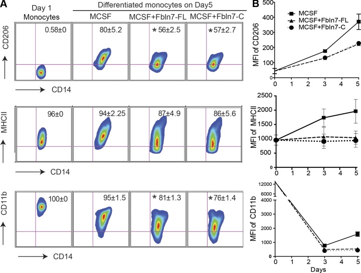
Fbln7-FL and Fbln7-C inhibit the differentiation of monocytes. Monocytes were cultured for 5 d in the presence of M-CSF (25 ng/ml), M-CSF + Fbln7-FL (10 μg/ml), and M-CSF+Fbln7-C (10 μg/ml), as described in Materials and Methods. A) Representative fluorescence-activated cell sorting plots from 3 independent experiments show the surface expression of CD206, MHC-II, and CD11b on FVS450−CD14+ macrophages at d 5. B) Line diagram represents mean fluorescence intensities (MFIs) of respective surface markers on monocytes that were cultured in the presence of M-CSF (solid line with square), M-CSF + Fbln7-FL (triangle broken line with triangle), and Fbln7-C (dotted line with circle) at d 3 (average of 4 separate experiments) and d 5 (average of 3 separate experiments). Results are expressed as means ± sem. *P < 0.05.

