
FIGURE 1.
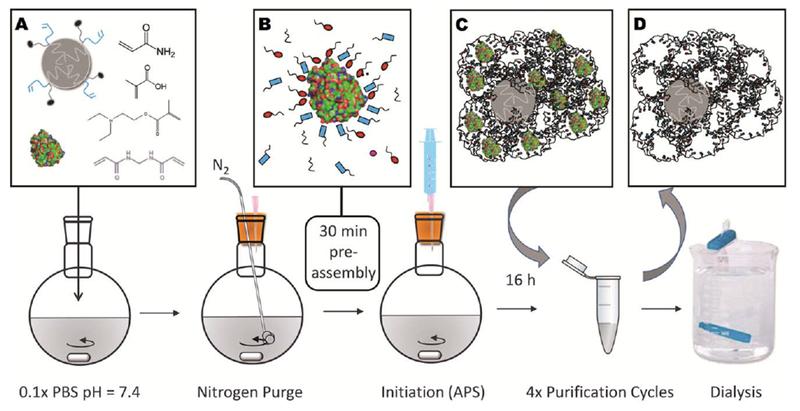
(a) PCL-NP, template protein, and functional monomers were dissolved in phosphate buffer and were (b) allowed to self-assemble for 30 min to form complementary hydrostatic and electrostatic interactions. In the graphic, the red ovals and blue rectangles represent the anionic, cationic, or otherwise functional moieties, which are contributed by each monomer. (c) Polymerized microparticles contained entrapped template, which was extracted with four purification cycles using 10% acetic acid, PBS, and ultrapure water, (d) leaving MIPs with void nanocavities. Dialysis was utilized for solvent exchange into water prior to lyophilization.