
FIGURE 2.
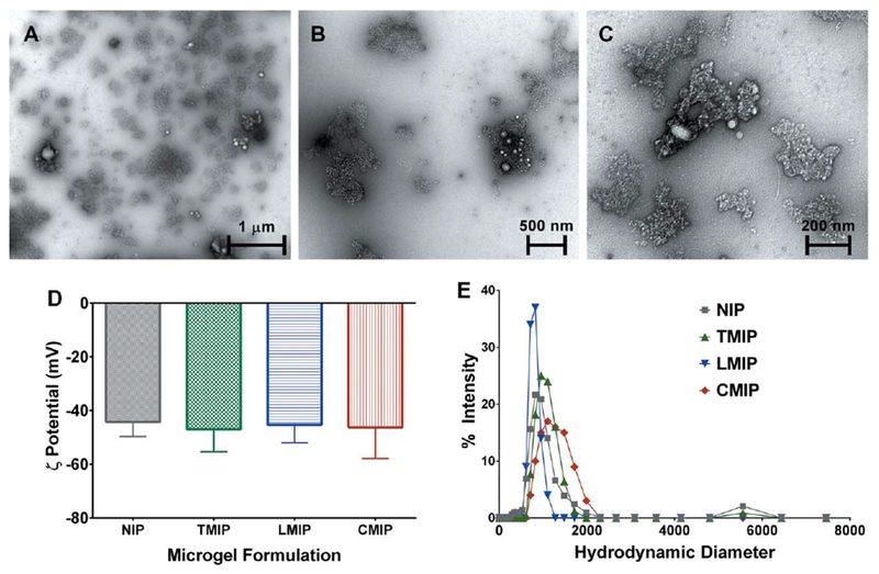
(a-c) Representative TEM images for imprinted and non-imprinted microgels. Imaging revealed that microparticles were highly polydisperse, irregularly shaped, and contained a varying number of PCL-NPs. LMIP shown, at increasing magnification to illustrate (a) the general distribution of microparticles, (b) typical microgel morphology, and (c) PCL-NP incorporation. No morphological differences were observed between MIPs and NIPs. (d) Zeta potential and (e) hydrodynamic diameter measurements were obtained at a microparticle concentration of 0.5 mg/mL in ultrapure water. The imprinting template identity did not significantly alter the microparticle z-average diameter or zeta potential. Quantitative metrics are presented in Table III. n = 3, presented as (d) zeta potential ± zeta deviation, and (e) average intensity.