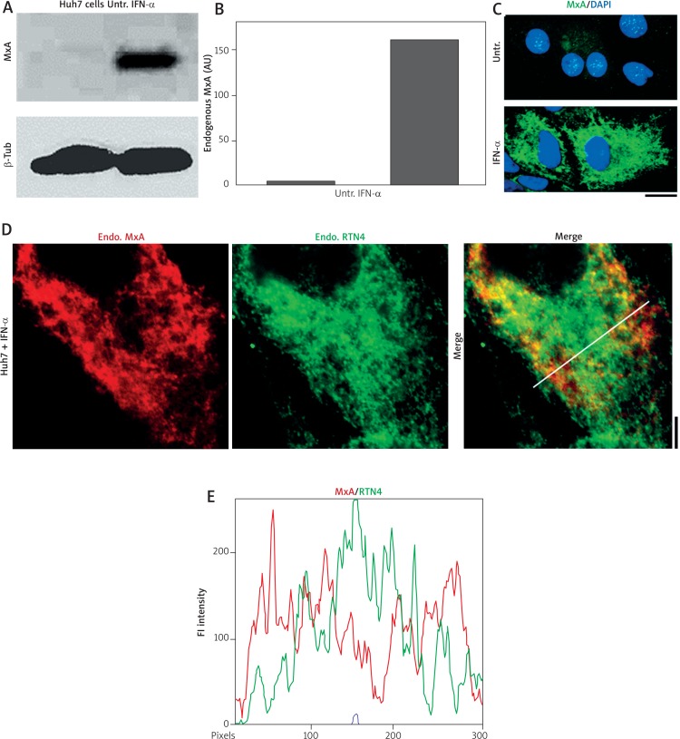
Fig. 1.
IFN-α-induced endogenous MxA in cytoplasm of Huh7 cells associated with structures distinct from the RTN4-based standard ER. Replicate cultures of Huh7 cells in 35 mm plates were exposed to recombinant IFN-α2a (3,000 IU/ml) in DMEM supplemented with 2% fetal bovine serum (“low-serum medium”) for 2 days or left untreated (Untr). Respective cultures were processed for preparing total cell extract or for double-label immunofluorescence imaging as indicated. Panels A and B, Western blots showing strong expression induction of MxA in Huh7 cells and quantitation (estimate: ~40-fold induction in this experiment). In the Western blots, β-tubulin was used as a loading control. Panel C, immunofluorescence images showing IFN-stimulated expression of endogenous MxA in the cytoplasm of Huh7 cells in a reticular pattern (25-35% of cells showed this pattern) imaged using a 40 × water-immersion objective. Scale bar = 10 μm. Panel D, Double-label immunofluorescence analyses of IFN-treated Huh7 cells for MxA and RTN4 imaged using an 100× oil immersion objective. Scale bar = 5 μm. The white line in the merged image in this panel indicates region depicted in the line scan in Panel E. R value indicated in the merged image in Panel D corresponds to the respective Pearson’s R coefficients (after automatic Costes’ thresholding) (co-localization corresponds to R > 0.8 [28])

