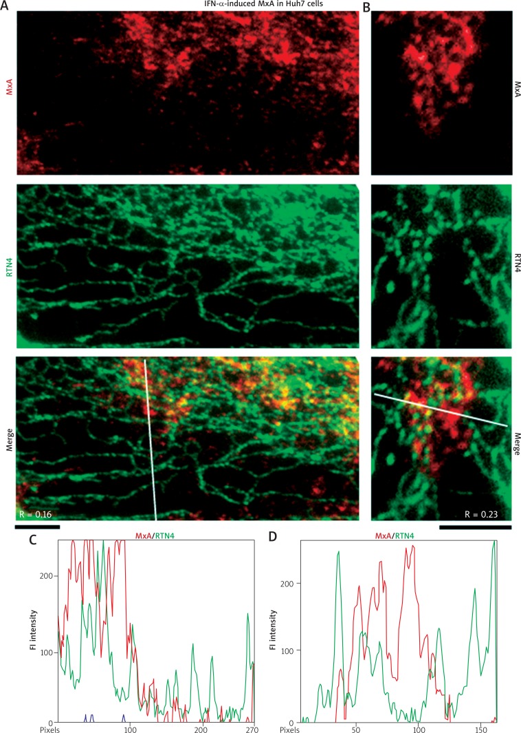
Fig. 2.
IFN-α-induced endogenous MxA in cytoplasm of Huh7 cells associated with structures distinct from RTN4 tubules of the standard ER. Replicate cultures of Huh7 cells in 35 mm plates were exposed to recombinant IFN-α2a (3,000 IU/ml) in DMEM supplemented with 2% fetal bovine serum (“low-serum medium”) for 2 days or left untreated (Untr) (as in Fig. 1), fixed and processed for double-label immunofluorescence imaging by sequentially probing for RTN4 first (in green) and then endogenous MxA (in red). The cells were imaged using an 100x oil immersion objective. Panels A and B represent two independent experiments. Scale bars = 5 μm. R values indicated in the merged images in Panels A and B correspond to the respective Pearson’s R coefficients (after automatic Costes’ thresholding). White lines in the merged images in Panels A and B indicate regions depicted in the line scans in Panels C and D respectively

