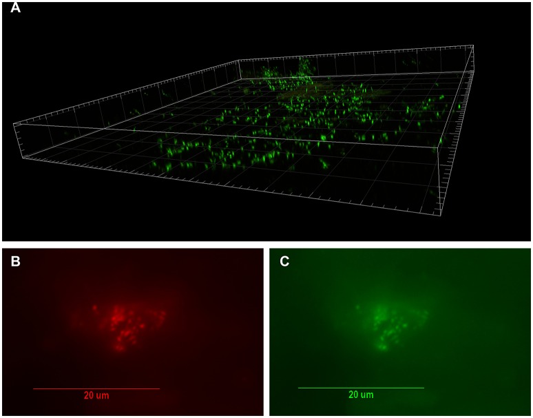
Figure 1.
Visualization of bacterial biofilm in the disc tissue by confocal laser scanning microscopy (CSLM) and confirmation of P. acnes by fluorescence in-situ hybridization (FISH) (reprinted from Capoor et al., 2017). (A) Three dimensional reconstructed CSLM image of biofilm bacteria stained with a DNA stain (SYTO9, green) in a disc tissue sample. (B,C) The presence of P. acnes biofilms in this sample verified using FISH. Epifluorescence micrographs of a biofilm cluster showing red fluorescence from the CY5-labeled EUB338 general eubacterial probe (B) and green fluorescence from the CY3-labled P. acnes-specific probe (C) Co-localization of the red and green fluorescence indicates that all of the bacteria in this biofilm were P. acnes.

