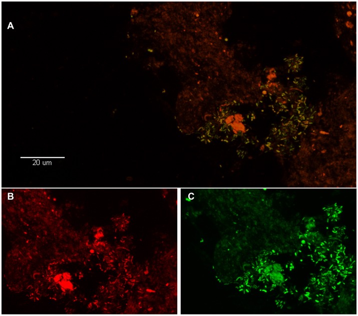
Figure 2.
Visualization of P. acnes biofilm in the disc tissue by use of FISH (reprinted from Capoor et al., 2017). (A) This color-combined image shows the “pocket” of green fluorescent P. acnes cells (biofilm) near the center right of the image. The presence of P. acnes biofilms in this sample was verified using FISH. (B,C) Red fluorescence is the general eubacterial probe (B) and green is the P. acnes probe (C). The (B,C) image is a zoom of (A) showing fluorescence from the red and green channels separately. Almost all of the cells in (A) are emitting both red and green fluorescence, indicating that they are P. acnes.

