. 2017 Aug 17;8(10):7014–7024. doi: 10.1039/c7sc01054k

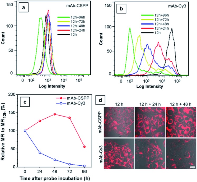
Fig. 8. Flow cytometric analysis of HCC827 cancer cells incubated with 10 μg mL–1 of (a) mAb–CSPP and (b) mAb–Cy3 at different time intervals. (c) Plot of relative fluorescence intensity (MFI/MFI12 h), where MFI12 h is the mean fluorescence intensity at 12 h incubation. Conditions: for mAb–Cy3, λex = 561 nm, detection with bandpass filter of 583 ± 7.5 nm. For CSPP, λex = 405 nm, detection with bandpass filter of 610 ± 10 nm. (d) The merged confocal images of the bright field and fluorescence images of cells incubated with mAb–dye conjugates for 12 h, and cells further incubated in fresh medium for 24 and 48 h. Conditions: for mAb–Cy3, λex = 560 nm, emission filter = 563–700 nm. For mAb–CSPP, λex = 405 nm, emission filter = 550–700 nm. Scale bar: 20 μm.

Fig. 8