Fig. 2.

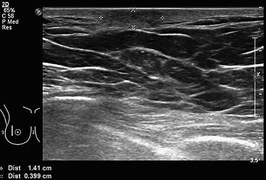
Patient A. Ultrasound: focal skin thickening of 5 mm (normal cutis is 2 mm). Diffuse hypoechogenic lesion with diffuse boundaries.

Patient A. Ultrasound: focal skin thickening of 5 mm (normal cutis is 2 mm). Diffuse hypoechogenic lesion with diffuse boundaries.