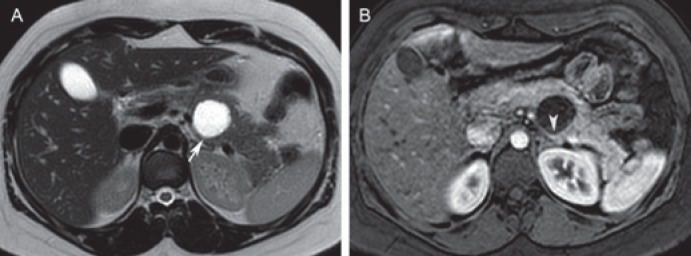
Fig. 4.
Mucinous cystic neoplasm in a 30-year-old female. A Axial T2-weighted image depicts a unilocular cystic lesion in the pancreatic body (arrow). The lesion is round and well circumscribed, and contains simple fluid. B Axial post-contrast T1-weighted image shows enhancement of a slightly thickened wall (arrowhead). Differential diagnosis includes an inflammatory cystic lesion. The patient did not have a history of pancreatitis, and the lesion was resected. On pathologic examination, this lesion proved to be a mucinous cystic neoplasm without dysplasia.

