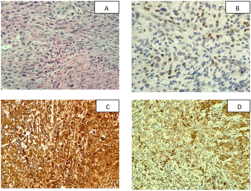
Figure 2. Histological analysis and immunohistochemical staining of the lung biopsy.
(A) Hemotoxylin and eosin staining showed that the tumor cells are predominantly composed of spindle cells with increased mitotic activity and areas of necrosis; (B) Tumor cells were weakly positive for P63 staining; (C) Vimentin showed strongly positive tumor cells; (D) Cytokeratin-Oscar showed tumor cells to be strongly positive

