
Figure 1.
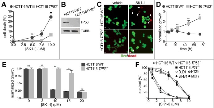
SK1-I decreases cancer cell growth and survival in a TP53-dependent manner. (A-C) Wild-type and TP53−/− HCT116 cells were treated with vehicle or with the indicated SK1-I concentrations for 24 h and the percentage of dead cells determined by live-dead assays. n = 4. (B) Immunoblots with the indicated antibodies for cells in panel (A). (C) Representative images of cells treated with 10 µM SK1-I for 24 h: live, green; dead, red. White arrowheads point to dead cells. (D-F) Wild-type and TP53−/− HCT116 cells were treated without or with 10 µM SK1-I for the indicated times (D) or with the indicated concentrations of SK1-I for 72 h (E) and cell proliferation determined. n = 3. (F) Wild-type, TP53−/− and P21−/− HCT116, DLD (mutant TP53), HT29 (mutant TP53), MCF7 (wild-type TP53), and BT474 (mutant TP53) cells were plated as single cell suspensions in 60-mm dishes and cultured 10 d for clonogenic assays. Colonies were fixed, stained, and colonies of >50 cells were counted. Survival data are expressed as percentage of colonies formed for each cell type treated with vehicle. n = 3. In (A,D,E,F) data are mean ± SEM; ns, not significant; *p ≤ 0.05; **p ≤ 0.005; #p≤ 0.0005. Scale bars: 10 µm.