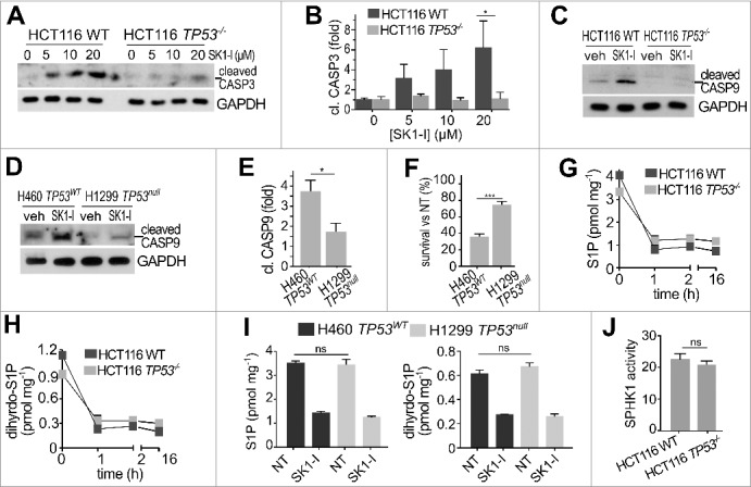
Figure 2.

Inhibition of SPHK1 with SK1-I induces cleavage of CASP3 and CASP9 in a TP53-dependent manner. (A-C) Wild-type and TP53−/− HCT116 cells were treated with vehicle or with increasing concentrations of SK1-I for 12 h (A,C) and analyzed by immunoblotting with the indicated antibodies, and cleaved CASP3 quantified by densitometry (B). n = 3. (D,E) H460 lung cancer cells with wild-type TP53, and H1299 TP53 null lung cancer cells were treated with vehicle or with SK1-I for 24 h, analyzed by immunoblotting with the indicated antibodies (D), and cleaved CASP9 quantified (E). n = 3. (F) Survival of H460 and H1299 cells was evaluated in clonogenic assays with 10 µM SK1-I. Cells were plated as single cell suspensions in 6-well dishes and cultured 10 d. Colonies were fixed, stained, and colonies of >50 cells were counted. Survival data are expressed as percentage of colonies formed for each cell type treated with vehicle (NT, not treated). n = 3. (G-I) LC-ESI-MS/MS analysis of S1P and dihydro-S1P in the indicated cell lines treated with 10 µM SK1-I for the indicated times (G,H) or for 2 h (I). Units are lipid per mg protein. (J) SPHK1 activity in wild-type and TP53−/− HCT116 cell extracts was measured with NBD-sphingosine as substrate.60 Data are mean ± SEM. n = 3. *p ≤ 0.05; ns, not significant.
