
Figure 6.
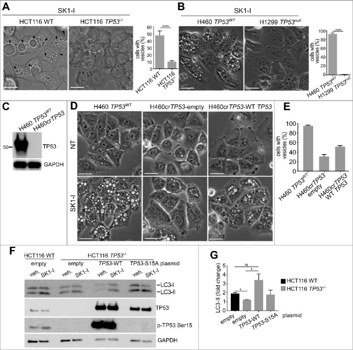
TP53 is required for SK1-I-induced accumulation of enlarged vacuoles and autophagy. (A, B) Phase contrast images and quantification of TP53WT and TP53−/− HCT116 cells (A), or H460 cells with wild-type TP53 and H1299 TP53-null cells (B), treated with 10 µM SK1-I for 1 h. (C) Immunoblot of H460 and CRISPR-Cas9-deleted TP53 H460 cells (H460crTP53). (D) Phase contrast images of H460, H460crTP53 cells transfected with an empty plasmid, or H460crTP53 cells transected with a plasmid expressing wild-type TP53, treated with vehicle or 10 µM SK1-I for 2 h. (E) Quantification of vesicle-positive cells in panel (D). (F,G) HCT116 wild-type cells transfected with empty vector, and HCT116 TP53−/− cells transfected with empty vector, or plasmids encoding wild-type TP53, or the TP53S15A mutant treated with 10 µM SK1-I for 12 h. Cell lysates were immunoblotted with anti-LC3 or the indicated antibodies. (K) Densitometric analysis of LC3-I and LC3-II shown in panel (J) n = 3. Scale bars (A, B, D): 10 µm.