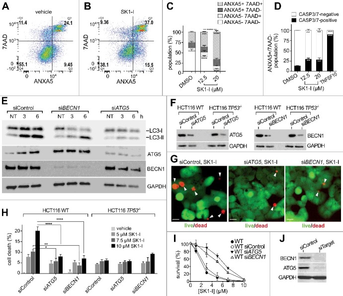
Figure 8.

Depleting BECN1 or ATG5 reduces SK1-I cytotoxicity. (A, B) Representative flow cytometry analysis of wild-type HCT116 cells treated with the vehicle (A) or 12.5 µM SK1-I (B) for 24 h followed by ANXA5 and 7-AAD staining. (C) Flow cytometric analysis of cells treated with the indicated concentrations of SK1-I for 24 h and labeled with CellEvent TM CASP3/7 Green for the final 6 h of SK1-I treatment followed by ANXA5 and 7-AAD staining. (D) Percentage of ANXA5+ 7-AAD- cells containing activated CASP3 and CASP7 in panel (C). TNFSF10/TRAIL-treated (100 ng/mL for 6 h) HCT116 wild-type cells were included as a positive control. (E) Wild-type HCT116 cells transfected with scrambled siRNA (siControl), siRNA targeting ATG5 (siATG5), or siRNA targeting BECN1 (siBECN1) were treated with 10 µM SK1-I for 20 h and cell lysates immunoblotted with the indicated antibodies. (F-H) HCT116 wild-type (F-H) and TP53-/- null (F, H) cells transfected with the indicated siRNAs were treated with 10 µM SK1-I (F, G) or with the indicated SK1-I concentrations for 12 h (H). n = 3. (G,H) Representative live or dead images (G) and live or dead quantification (H). White arrowheads in (G) point to dead cells. (I) Clonogenic assay of wild-type HCT116 cells transfected with siControl, siATG5, or siBECN1 treated with vehicle or the indicated SK1-I concentrations, cultured for 10 d, and colonies of >50 cells counted. n = 3. (J) Cell lysates of duplicate cultures in panel (I) were immunoblotted with the indicated antibodies (F-I). n = 3, mean ± S.D. ns, not significant; *p≤0.05; **p≤0.005; ***P≤0.001;****P≤0.0005.
