Figure 1.

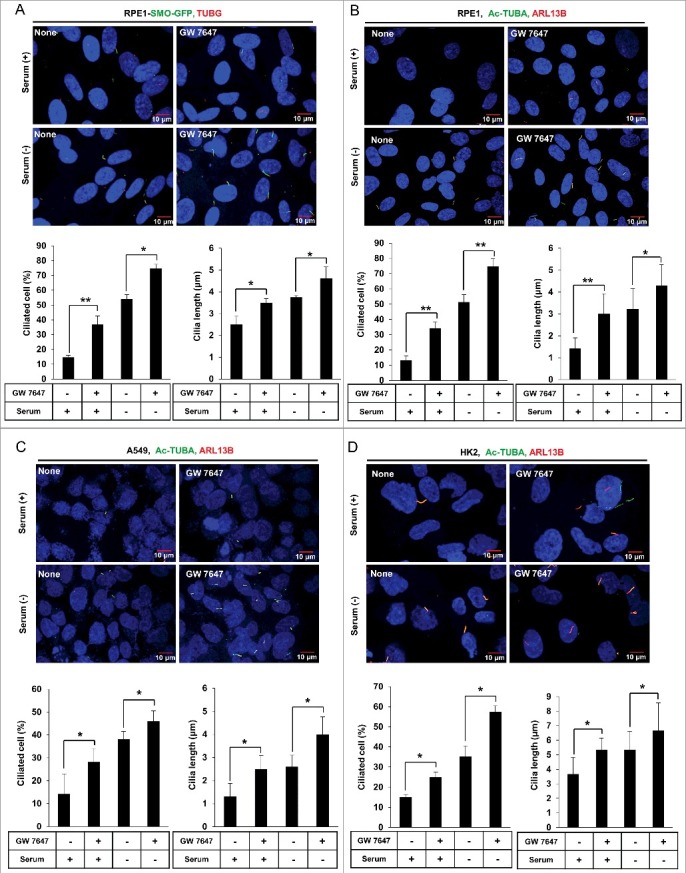
The PPARA ligand promotes ciliogenesis in mammalian cells. (A) RPE1-SMO-GFP cells were treated with 0.5 μΜ GW 7647 as indicated. After incubation for 24 h, cells were immunostained with anti-TUBG Alexa Fluor 568-conjugated antibody and observed using fluorescence microscopy. Merged signal showed the primary cilium. The number of ciliated cells and the length of cilia were quantified as described in the materials and methods section. (B, C, and D) RPE1, A549, and HK2 cells were treated with 0.5 μΜ GW 7647 as indicated. After 24 h, cells were immunostained with anti-Ac-TUBA Alexa Fluor 488-conjugated antibody and anti-ARL13B Alexa Fluor 568-conjugated antibody. The cells were observed using fluorescence microscopy. The primary cilium was showed by merged signal. The number of ciliated cells and the ciliary length of the cells were quantified as described in the materials and method section. All data shown represent mean ± s. d. percentage of cells with primary cilia or length of cilia for 200 cells per well in triplicate samples. *P < 0.05, **P < 0.01 and ***P < 0.001, one-way ANOVA, as compared to that of cells incubated in normal or serum-free media only.
