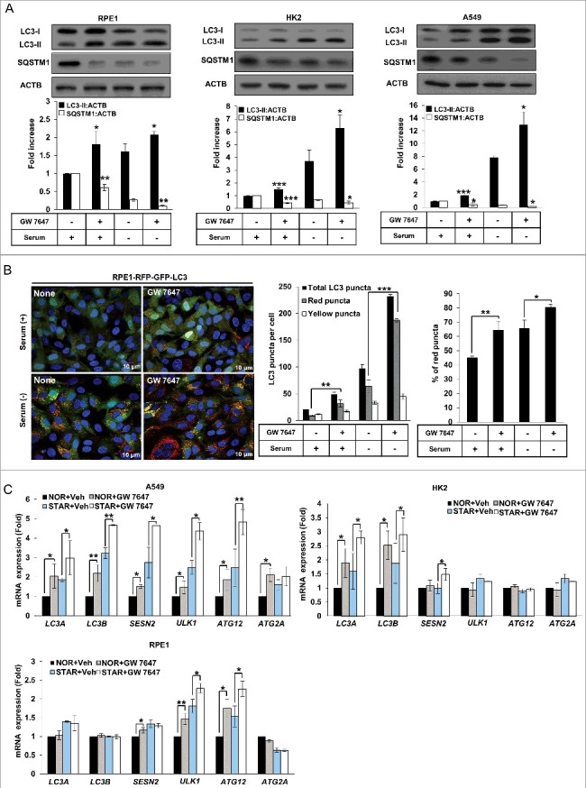
Figure 2.

The PPARA ligand upregulates autophagy in mammalian cells. (A) RPE1, HK2, and A549 cells were treated with 0.5 μM GW 7647 as indicated. After 24 h, whole extracts were separated by SDS-PAGE and western blot analysis was conducted with antibodies against LC3, SQSTM1, and ACTB. (B) RPE1-RFP-GFP-LC3 cells were treated with 0.5 μΜ GW 7647 as indicated. After incubation for 24 h, cells were fixed with 4% paraformaldehyde, and mounted with DAPI. The number of red LC3 puncta and yellow LC3 puncta per cell in each condition was quantified. Total puncta are the sum of the number of red and yellow puncta. Percentage of the red puncta is from the total LC3 puncta. Data shown represent mean ± s. d. number of red and yellow puncta for 50 cells per well. (C) RPE1, HK2, or A549 cells were treated with vehicle (Veh) or 0.5 μM GW 7647 as indicated. After incubation for 12 h, total RNAs were extracted, and expression of LC3A, LC3B, SESN2, ULK1, ATG12, and ATG2A was analyzed with quantitative real-time polymerase chain reaction (qPCR). NOR: normal; STAR: serum starvation. All the experiments were performed 3 times. *P < 0.05, **P < 0.01 and ***P < 0.001, one-way ANOVA, compared to cells incubated in normal or serum-free media only.
