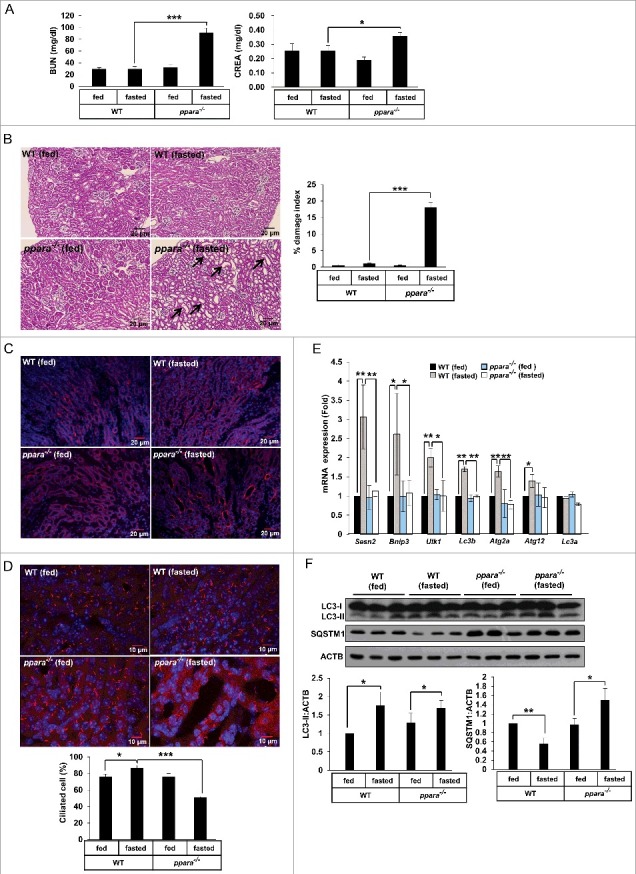
Figure 6.

PPARA deficiency induces kidney damage and loss of cilia through impaired autophagy. (A) Blood samples from chow-fed wild-type (WT) mice, 48 h fasted WT mice, chow-fed ppara−/− mice, and 48 h fasted ppara−/− mice (n = 6) were prepared and serum levels of creatinine (CREA) and blood urea nitrogen (BUN) were determined. (B) Kidneys from chow-fed WT mice, 48 h fasted WT mice, chow-fed ppara−/− mice group, and 48 h fasted ppara−/− mice (n = 6) were removed and embedded in paraffin, and 5 μm sections were prepared. Kidney specimens were stained with hematoxylin and eosin (H&E). Damaged indices were calculated based on representative renal sections as described in Materials and Methods. (C) Kidney specimens (n = 6) were stained with Dolichos biflorus agglutinin (DBA, red). (D) Kidney specimens (n = 6) were immunostained with anti-ARL13B and Alexa Fluor 568-conjugated antibody as indicated. Data shown represent mean ± s. d. percentage of cells with primary cilia for 1000 cells per kidney in 6 samples. (E and F) Total RNAs or proteins were extracted from kidneys (n = 6) and analyzed by quantitative real-time polymerase chain reaction (qPCR) or immunoblotted as indicated. Black arrows, damaged area. *P < 0.05, **P < 0.01 and ***P < 0.001, one-way ANOVA, as compared to those from WT fasted group or WT fed group.
