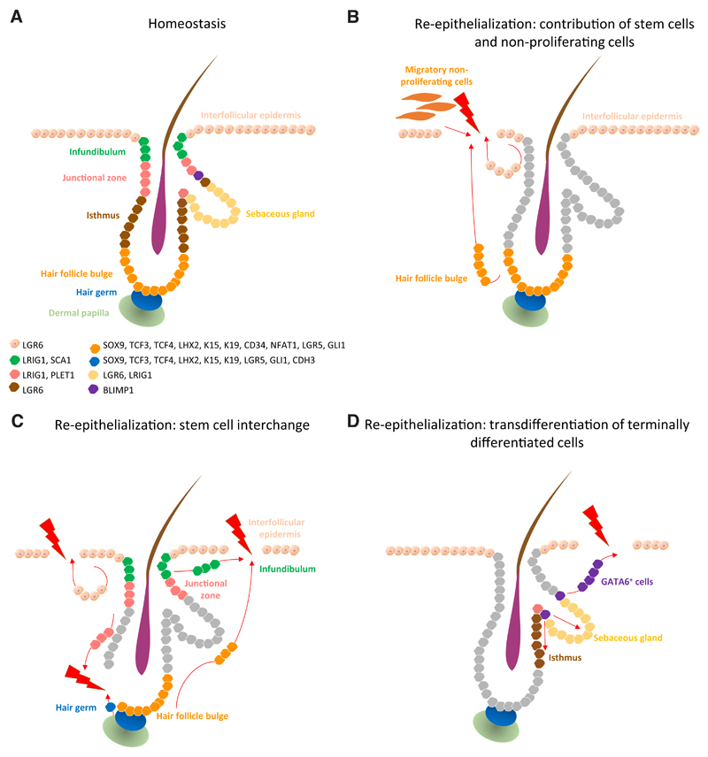
Fig. 4. Mechanisms of re-epithelialization.
Epidermal stem cell compartments that maintain skin homeostasis and their associated markers (A). Re-epithelialization upon injury occurs via several paths: contribution of the proliferative hub (IFE hair- follicle stem cells and their progeny) and non-proliferative migratory cells (at the leading edge) to the initial stages of re-epithelialization (B). When stem cell compartments from the IFE, infundibulum, junctional zone and hair follicle bulge and germ exhibit plasticity, they contribute to the replenishment of stem cells lost on wounding (C). Terminally differentiated cells such as GATA6+ cells de-differentiate and contribute to re- epithelialization of damaged IFE and re-populate the sebaceous gland and lower hair follicle during wound healing (D).

