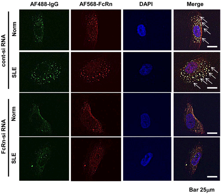
Figure 2.
IgG entry into podocytes via the neonatal Fc receptor (FcRn). Localization of IgG derived from a normal subject (Norm) and a patient with systemic lupus erythematosus (SLE) nephritis (Alexa Fluor [AF] 488 stained) in podocytes was analyzed by immunofluorescence staining, with or without silencing of FcRn (Alexa Fluor 568 stained), the receptor of IgG, using FcRn small interfering RNA (siRNA) or control siRNA. Arrows indicate entry of IgG into the cytoplasm, which did not occur when FcRn was silenced.

