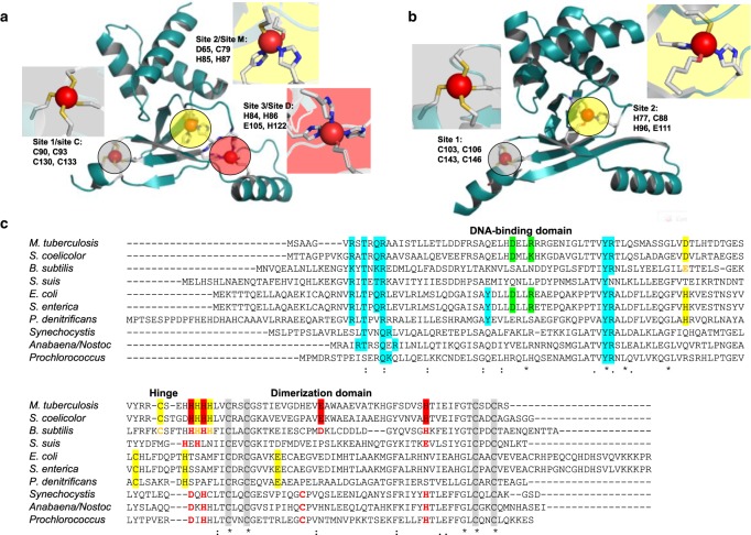
Figure 5. Structural and sensory zinc sites on Zur proteins.
(A) ScZur (pdb 3mwm [38]) and (B) EcZur (pdb 4mtd [17]). The structural sites are highlighted in grey, the single or major sensory site in yellow, and the additional site in ScZur is highlighted in red. (C) Sequence alignment of Zur proteins from a variety of species. Residues confirmed to participate in zinc binding by X-ray crystallography are highlighted by red, yellow, and grey backgrounds. Residues involved in DNA binding are highlighted in cyan. Predicted metal-binding residues or sensory sites in Zur proteins that have not been structurally characterised are printed in red or yellow. The two residues forming a salt bridge in EcZur (see the text) are highlighted in green; they are (semi-)conserved in Zur from Salmonella, M. tuberculosis, and S. coelicolor.

