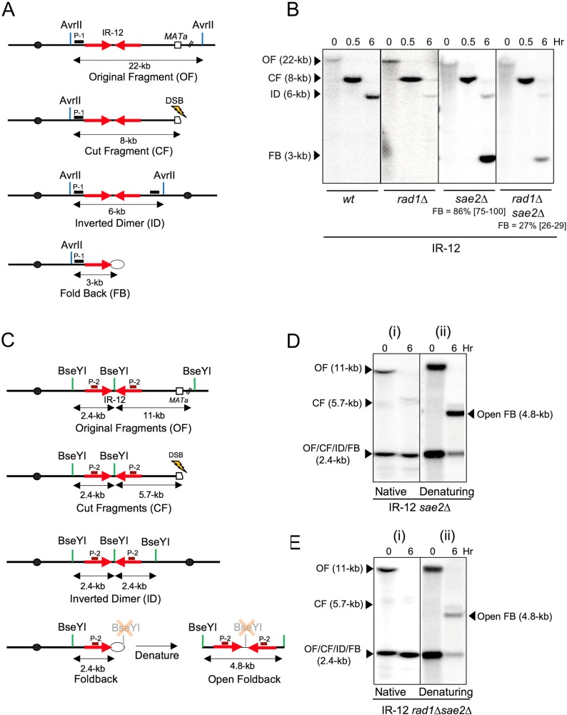
Fig 4. Inverted dimers and fold-backs in IR-12.
(A) Schematics of AvrII- digested Chr III in IR-12 (OF) and its derivatives: CF, ID, FB (location of probe P-1 is indicated by black box). (B) DSB repair in IR-12 (wt, rad1Δ, sae2Δ and rad1Δsae2Δ) following AvrII digest and hybridization to probe P-1. The respective positions of, CF, ID and FB are indicated. The median efficiencies of FB formation (%) and the range of the median [in brackets] are indicated (see S1 Fig and Materials and Methods for details). (C) The schematics of BseYI restriction fragments of Chr III in original IR-12 and its derivatives following DSB: OF, CF, ID, FB and Open-FB (formed by denaturation of FB). The location of hybridization probe P-2 is indicated by brown box. (D) Southern blot analysis of DSB repair in sae2Δ derivative of IR-12 using native and denaturing gel electrophoresis (Schematics in Fig 4C) and hybridization to probe P-2. The positions and corresponding sizes of OF, CF, ID, FB and Open-FB following BseYI digestion of DNA isolated before (0-hr) and 6-hr following DSB induction are indicated. (E) Southern blot analysis of DSB repair in rad1Δsae2Δ derivative of IR-12 using native and denaturing gel electrophoresis (schematics in Fig 4C) following hybridization to probe P-2.

