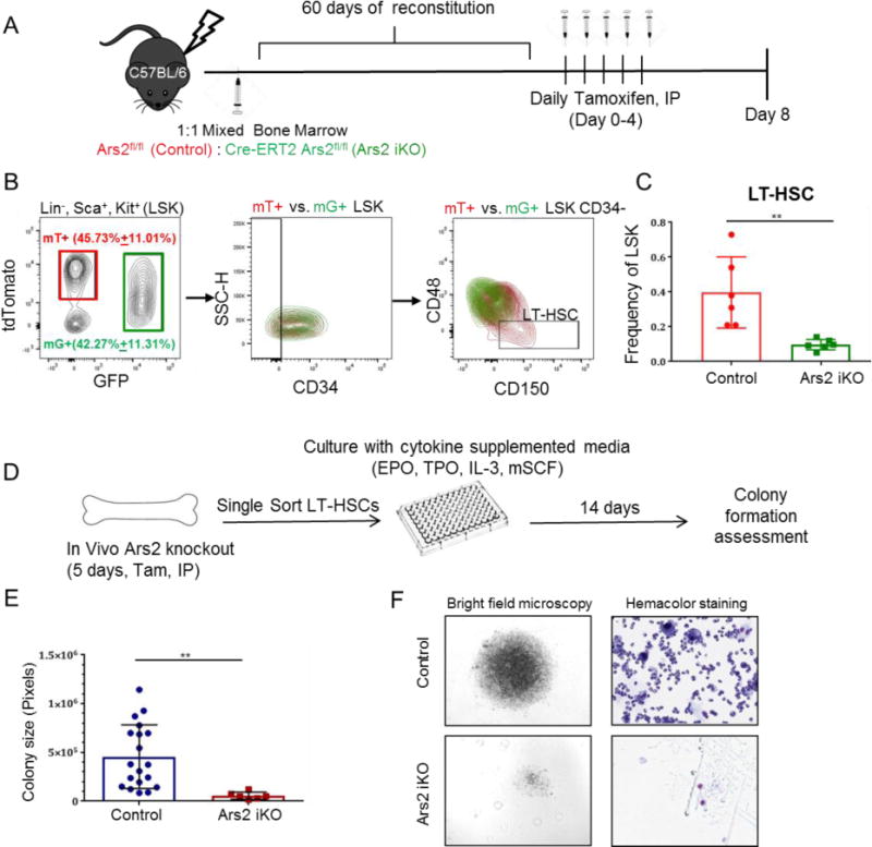
Figure 6. Defect in Ars2 knockout LT-HSCs is cell-intrinsic.

A) Schematic of mixed mT/mG chimera generation. Two months after reconstitution with 1:1 bone marrow cells from both Cre+ mT/mG and Cre- mT/mG mice, mice were injected with daily tamoxifen and bone marrow was analyzed at day 8. B) Representative density plots with mT+, wild-type (red) and mG+, Ars2 knockout (green) cells isolated from a single mixed chimera overlaid to demonstrate gating strategy and intrinsic differences between cell populations. C) Frequency of Ars2 knockout, mG+ LT-HSCs (green bar) was reduced compared to wild-type, mT+ LT-HSCs (red bar) in mixed chimeras eight days following initial tamoxifen injection. n=6, **p=0.002. D) Schematic of single cell colony formation assay. Single LT-HSCs from tamoxifen-treated control or Ars2 iKO chimeras were FACS sorted into 96 well plates and cultured for 14 days in cytokine supplemented media. E) Size of colonies formed from Ars2 iKO LT-HSCs was significantly less than size of colonies formed from control LT-HSCs (**p = 0.007). Each dot represents a colony. F) Comparison of representative colony size (left) and differentiation (right) obtained from a singly sorted LT-HSC from a tamoxifen-treated control chimera (top) versus a tamoxifen-treated Ars2 iKO chimera (bottom).
