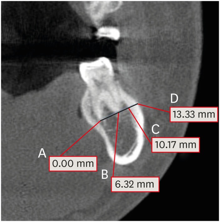
Figure 2. Representative measurements on the coronal plane of a cone-beam computed tomography image. Four points that intersected with the horizontal line crossing the apex of DB and DL root in a mandibular first molar with the morphology of 2 separate distal roots with 1 canal in each root were set up, as follows: (A) DL root apex; (B) DB root apex; (C) inner surface of the buccal cortical bone; (D) outer surface of the buccal cortical bone. The measurements at 4 points are the distances from point A.

DB, distobuccal; DL, distolingual.
