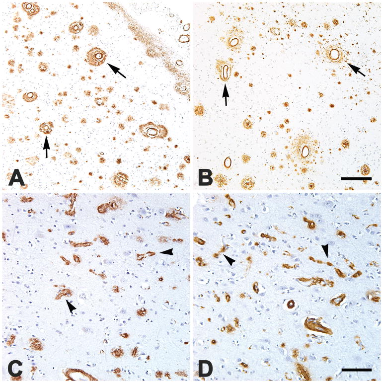
FIGURE 1.
Severe, dyshoric large-vessel CAA (A,B; four vessels are marked by arrows) in occipital (A) and parietal (B) neocortical sections, and severe dyshoric capillary CAA (C,D; four are marked by arrowheads) in the parietal neocortices of two African-Americans (A,C) and two Caucasians (B,D). Areas of heavy capCAA generally had relatively few parenchymal Aβ plaques (C,D). Nissl counterstain. Scale bar in B = 200 μm for A&B, scale bar in D = 100 μm for C&D.

