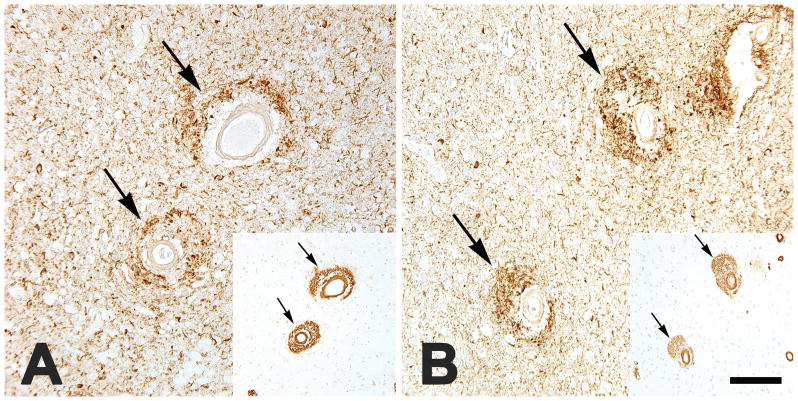
FIGURE 2.
Large cortical blood vessels with dyshoric CAA are surrounded by profuse neuritic tauopathy (antibody PHF-1), here shown in the parietal neocortex of an APOEe3/4-bearing African-American man (AD7; A) and the occipital neocortex of an APOEe3/3-bearing Caucasian woman (AD27; B). Insets show intramural and perivascular Aβ immunoreactivity in the same vessels in nearby tissue sections (antibody 4G8). Note the spatial overlap of tauopathic neurites and perivascular Aβ. Nissl counterstain. Bar = 200 μm for the insets, and 80 μm for the main panels.

