
Figure 3.
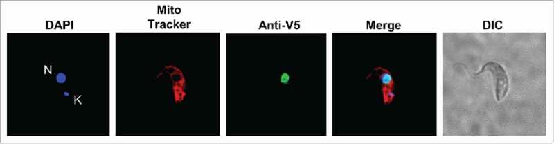
TbTGT is a nuclear enzyme. Immunofluorescence localization performed with cells expressing a V5-epitope tagged TbTGT. Anti-V5 antibodies were used to detect TbTGT. Mitotracker was used to stain the mitochondria (red) while DAPI stained the nuclear (N) and mitochondrial DNA (K) (blue). DIC refers to a phase-contrast image. The figure is representative of at least five different experiments.