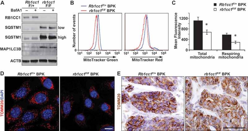
Figure 2.

Rb1cc1 deletion reduces mitochondrial mass in BPK mammary tumors. (a) Immuno-blots showing levels of RB1CC1, SQSTM1, MAP1LC3B and ACTB in BPK primary cells with Rb1cc1F/+ or rb1cc1F/F. Cells were treated with HBSS ± bafilomycin A1 at 200 nM for 2 h before harvesting lysates. On the right, low indicates lower exposure time and high indicates higher exposure time of the same blot. (b) Histograms showing the distribution of BPK primary cells stained with MitoTracker Green or MitoTracker Red and analyzed by flow cytometry. (c) Quantification of the mean fluorescence intensities of BPK primary cells stained with MitoTracker Green or MitoTracker Red. Data represent the average of n = 4 tumors for Rb1cc1F/+ and rb1cc1F/F cohorts. Statistical significance was determined by two-tailed t-test, * denotes p ≤ 0.05. (d) Representative images of BPK primary cells with Rb1cc1F/+ or rb1cc1F/F alleles that were immuno-fluorescently labeled for TOMM20 and analyzed via confocal microscopy. Scale bar: 10 μm. (e) Representative images of BPK tumors with Rb1cc1F/+ or rb1cc1F/F alleles that were immuno-stained for TOMM20. Scale bar: 200 μm.
