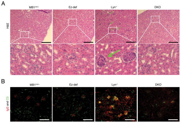
Figure 4. B cell-specific deletion of ezrin in Lyn−/− mice leads to inhibition of glomerulonephritis.
(A) H&E stained kidney sections from 8 month old MB1cre/+, Ez-def, Lyn−/− and DKO mice. Insets marked by white boxes show more highly magnified glomerular areas showing leukocyte infiltration in Lyn−/− kidneys and lack thereof in MB1cre/+, Ez-def, and DKO kidneys. (B) Immunofluorescence images of kidneys from 8 month old MB1cre/+, Ez-def, Lyn−/− and DKO mice stained with antibodies to IgG (red) and complement fragment C3 (green). Data represent at least four mice of each genotype in two independent experiments. Scale bars, 50 μm.

