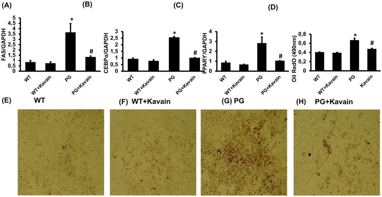
Figure 3.
Effect of Kavain treatment on 3T3-L1 derived adipocytes cells with and without exposure of P. gingivalis. mRNA expression of adipogenic markers (A) FAS, (B) CEBPα, (C) PPAR-γ, (D) Oil Red O absorbance measured at 490nm and lipid droplets formation (E, F, G&H) pictures presented lipid accumulation after Oil Red O staining in WT-control, WT+Kavain, P.gingivalis exposed adipocytes and P.gingivalis + Kavain exposed cells. Results are mean±SE, n=3–4, *p<0.05 vs. WT, #p< 0.05 vs. P.gingivalis.

